 |
FEATURED ITEMS |
 |
|
|
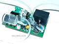
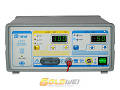
| CV-2000A 2004 Argon Plasma Coagulation Unit
|
|
|
CV-2000A 2004 Argon Plasma Coagulation Unit, is equal parts cutting, coagulation, bipolar, and argon ion coagulation. It can satisfy the demands of different surgeries. With a endoscopic operating modes, the output of argon gas is less than 0.2L/ min.
product number: 2004
Click for More Details
|

|
|
|
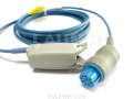
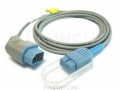
| NT3B Multparameter Patient Monitor
|
|

|
NT3B Multparameter Patient Monitor,12.1" High Brightness TFT LCD,1-8 Channel Waveform Display,7 Channel ECG Waveform Display; ST Segment Analysis; Arrhythmia Analysis,Anti-movement and low perfusion SpO2 Measurement,Defibrillation and ESU Protection.
Click for More Details
|
|
|
|
|
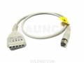
| IBP Module B300X MOCT-B300X the external version IBP
|
|
|
The IBP Module Model B300X, the external version, uses PLUG & PLAY IBP TECHNOLOGY with dual-channel measurement. The IBP Module also features low-power, no extra power supply is needed,
automatic digital data processing, and can repeatedly zeroing, calibration function
and can be used to monitor ART, PAP, CVP, RAP, LAP, ICP and P1-P2
Click for More Details
|
|
|
|
|


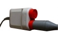
 Emergency and Clinics
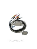
 Auto Diagnostic Cable